

Biofabrication & Clinical Applications of Biomaterials in Biomedical Engineering
Biomaterials in medical devices / 3D bioprinting and biofabrication
Biofabrication & Clinical Applications of Biomaterials
Biofabrication platforms are becoming central to next-generation medical device design
3D bioprinting, electrospinning, and decellularisation each address a gap that conventional biomaterials and fabrication routes struggle to solve. Together they enable spatially organised structures, extracellular-matrix-mimicking architectures, and biologically derived scaffolds that more closely reflect native tissue complexity.
The opportunity is substantial, but so is the translational challenge. These platforms increase the burden on process validation, regulatory strategy, and scale-up discipline, particularly where biological components, patient-specific design, or combination product rules come into play.
Platform novelty alone does not determine success. Development discipline does.
Building on FutureBridge perspective on bioprinting and personalised healthcare, this page positions biofabrication as the next strategic layer in biomaterials innovation when structure, biology, and patient-specific performance need to be engineered together.
Figures indicative; align with internal regulatory and market assumptions before publication.
Why biofabrication is essential in advanced medical device design
Advanced devices increasingly require more than bulk material performance. They need controlled architecture, tissue-like interfaces, local biological signalling, and in some cases patient-specific geometry. Biofabrication platforms address these requirements by shaping how biomaterials are organised in space, not just what those biomaterials are made of. For the broader context of active, stimuli-responsive, and regenerative material classes, see Active, Adaptive, and Regenerative Biomaterials are Creating New Growth Frontiers.
Clinical signal
- 3D bioprinting enables patient-matched geometry for skull, spine, and cartilage-related constructs.
- Electrospinning is already commercially mature for several acellular scaffold applications.
- Decellularised matrices preserve native architecture and signalling that synthetic scaffolds cannot fully replicate.
The most successful biofabrication programmes are not defined only by platform novelty. They are defined by how early teams lock classification, evidence design, scale-up assumptions, and clinical use-case fit.
Key biofabrication platforms
These three platforms represent distinct ways to engineer biological performance into devices and scaffolds. Their commercial maturity, translational risk, and manufacturing logic differ significantly, which is why platform selection should be tied to indication, evidence burden, and operating model from the outset.
3D bioprinting
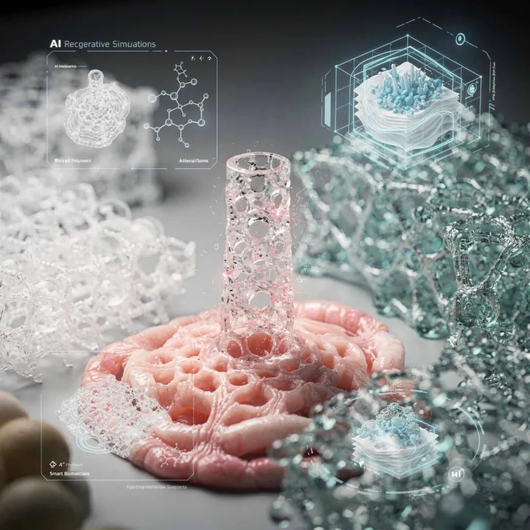
3D bioprinting builds three-dimensional architectures through layer-by-layer deposition of biomaterial inks, cell-laden bioinks, or acellular support structures. Extrusion handles high-viscosity hydrogels and polymer melts, inkjet supports low-viscosity bioinks with high resolution, and stereolithography uses photopolymerisation for complex geometries with high fidelity.
Clinical appeal: Patient-specific geometry, including skull implants matched to defects, spinal cages tuned to disc dimensions, and cartilage constructs that aim to replicate native zonal architecture. For the ceramic and HA-PLGA composite scaffold context, see Ceramic Biomaterials: Hydroxyapatite, Bioactive Glass and Bioceramic Implants.
Electrospinning
Electrospinning uses high-voltage electrical fields to draw polymer solutions into fibres ranging from tens of nanometres to several micrometres, producing non-woven mats that mimic extracellular matrix structure. Electrospun PLGA, PCL, and collagen scaffolds — covered in depth in Polymeric Biomaterials & Scaffolds: PLA, PLGA, PEEK and Beyond — are already used across wound care, vascular grafts, nerve conduits, and tendon repair concepts.
Clinical signal: Electrospun membranes are among the most commercially mature biofabricated formats for acellular scaffold applications, with multiple approved device pathways demonstrating repeatability and translational fit.
Decellularisation
Decellularisation removes cellular components from tissues or organs while preserving extracellular matrix architecture, vasculature, and biochemical signals. The result is a biological scaffold that retains growth factors, adhesion proteins, and structural proteins that synthetic materials cannot fully reproduce.
Clinical examples: Decellularised heart valves, small intestinal submucosa for wound and hernia repair, and bladder matrices for urological reconstruction; batch-to-batch variability remains the key manufacturing and regulatory challenge.
The 4-stage clinical translation pathway
The journey from a biofabricated concept to commercial approval follows four stages. Each stage has distinct evidence outputs, primary risks, and critical decisions. When these stages are compressed or treated as engineering handoffs instead of programme gates, timelines extend quickly.
Stage 1
Discovery & feasibility
Material system, fabrication route, and target indication are defined. In-vitro testing establishes biocompatibility against the relevant ISO 10993 exposure category, while initial mechanical testing characterises scaffold or device performance.
Key action: Lock device classification and begin pre-submission dialogue early; misclassification here can add years later.
Stage 2
Design & development
Device design is optimised for clinical performance, sterilisation, shelf-life, and manufacturing scalability. For biofabricated devices this includes bioink formulation lock, process development, sterilisation validation, and packaging design.
Key action: Treat design lock and ISO 10993 programme design as one integrated activity; late design changes are schedule events, not minor iterations.
Stage 3
Evidence generation
Animal studies and, for higher-risk devices, clinical investigations generate the safety and performance evidence required for submission. Novel biofabrication platforms often move into advanced evidence pathways where equivalence is hard to defend.
Key action: Design clinical investigations with regulatory and HTA endpoints in view, and run payer and regulator engagement in parallel.
Stage 4
Submission & launch
Submission consolidates evidence from the previous stages. Where biological components or drugs are integrated, combination product rules can shape review leadership, dossier structure, and approval timelines. Launch readiness also depends on training, surveillance, and reimbursement planning.
Key action: Seek combination product designation early if a biological or drug component is present; that decision can materially shift timelines and review logic.
A framework to select the right biofabrication route
Platform selection should align fabrication capability with therapeutic intent, evidence burden, and operational readiness. The table below provides a practical screening view for R&D teams evaluating which route best fits the target application. For a broader framework on translating biomaterial innovation into commercial products, see How can your Biomaterial Innovation achieve commercial viability?.
| Platform | Primary Strength | Biological Complexity | Manufacturing Challenge | Regulatory Maturity | Nearest Commercial Application |
|---|---|---|---|---|---|
| 3D bioprinting | Patient-specific architecture and spatial control | High to very high | High | Emerging | Customised implants, scaffold constructs, regenerative platforms |
| Electrospinning | ECM-like fibre architecture | Low to moderate | Moderate | More mature | Wound dressings, membranes, acellular scaffolds |
| Decellularisation | Native biological structure and signalling | High | High due to donor variability | Selective but established in some applications | Heart valves, SIS-based repair matrices, bladder scaffolds |
Key pain areas in biofabrication programmes
The opportunity in biofabrication is significant, but the operating burden is equally real. Platform novelty, biological variability, and evidence design interact in ways that can reshape both development cost and time to market.
1. Classification errors distort the entire programme
Teams often underestimate how quickly a device concept can move into biologic or combination-product logic once cell-laden systems, bioactive agents, or tissue-derived matrices are introduced. That shift changes evidence expectations and review leadership.
Solution: Start regulatory mapping at feasibility stage and align platform, indication, and classification logic before design lock.
2. Scale-up undermines structural consistency
Bioink rheology, fibre morphology, porosity, and donor-derived matrix composition can all shift during scale-up. A construct that performs well at lab scale may behave differently under commercial process conditions.
Solution: Build in process analytics, formulation control, and manufacturing-transfer logic early instead of treating scale-up as a post-proof-of-concept exercise.
3. Evidence design often lags commercial reality
Programmes may generate technically sound data but still fail to support reimbursement, clinician adoption, or payer confidence if endpoints are not chosen with downstream decision-makers in mind.
Solution: Integrate clinical, regulatory, and HTA endpoints early so the evidence package supports both approval and market access. FutureBridge Navigating the Biomaterial Challenge sets out a structured framework for this decision.
FutureBridge perspective
Biofabrication is shifting from platform curiosity to MedTech strategy
Biofabrication is no longer just a technology watch topic. It is becoming a strategic decision area for companies working at the intersection of scaffolds, regenerative medicine, patient-specific implants, and advanced biomaterial architectures.
The real decision is not whether these platforms are promising. It is where they fit commercially, which indications justify the evidence burden, and how early-stage design choices can preserve optionality instead of locking teams into avoidable regulatory and manufacturing complexity.
Frequently asked questions
What is 3D bioprinting in medical device development?
3D bioprinting is a layer-by-layer fabrication method that deposits biomaterial inks, cell-laden bioinks, or support materials to create three-dimensional constructs with controlled spatial organisation. It is especially valuable where patient-specific geometry, structural gradients, or local biological functionality need to be engineered into the device or scaffold.
Why is electrospinning important for scaffold design?
Electrospinning creates fibre networks that resemble extracellular matrix architecture, making it highly relevant for wound care, tissue support, and acellular scaffold formats. Its importance lies in combining structural simplicity with clinically useful surface area, porosity, and fibre-scale control.
What makes decellularised scaffolds different from synthetic biomaterials?
Decellularised scaffolds preserve native extracellular matrix structure, signalling molecules, and biological cues that synthetic materials cannot fully reproduce. That gives them strong translational appeal in repair and reconstruction applications, but also introduces manufacturing variability tied to source tissue and processing consistency.
What is the biggest risk in translating biofabricated devices to market?
The biggest risk is usually not the platform itself but the mismatch between platform complexity and development planning. Classification, evidence design, process validation, and scale-up assumptions must be aligned early; otherwise programmes accumulate expensive delays that are difficult to recover later.